Are you curious about the orbicularis oculi muscle and how it helps you close your eyelids? Look no further! In this article, we’ll explore the fascinating anatomy, function, and origin of this essential eye muscle. The orbicularis oculi is responsible for closing your eyelids and aiding in tear drainage. Its two parts, the orbital section and the palpebral section, control both voluntary and involuntary closure of the eye. Join us as we uncover the secrets of this crucial muscle and its impact on eye health.
Anatomy of the Orbicularis Oculi
The anatomy of the orbicularis oculi muscle consists of three parts: the orbital, palpebral, and deep palpebral sections. The orbital section is responsible for the voluntary closure of the eyelids, while the palpebral section is involved in both voluntary and involuntary closure of the eye. The deep palpebral section plays a role in compressing the lacrimal sac. The muscle is innervated by the zygomatic and temporal branches of the facial nerve.
Understanding the anatomy of the orbicularis oculi muscle is crucial in identifying and treating disorders associated with its dysfunction. Conditions like Bell’s palsy or paralysis of the facial nerve can lead to poor closure of the eyelids, resulting in exposure keratitis and corneal problems if not addressed properly. Other disorders that can affect the muscle include blepharospasm, facial nerve injuries, aging-related changes, and medical conditions like myasthenia gravis.
Treatment options for orbicularis oculi muscle disorders may include botulinum toxin injections, surgical interventions, physical therapy exercises, and medications. Ongoing research and advancements in this field focus on the efficacy of different treatment modalities, understanding the underlying mechanisms and causes of muscle dysfunction, advancements in surgical techniques, and the development of targeted therapies. Collaborative efforts between researchers, clinicians, and patients contribute to advancements in the field of orbicularis oculi muscle disorders.
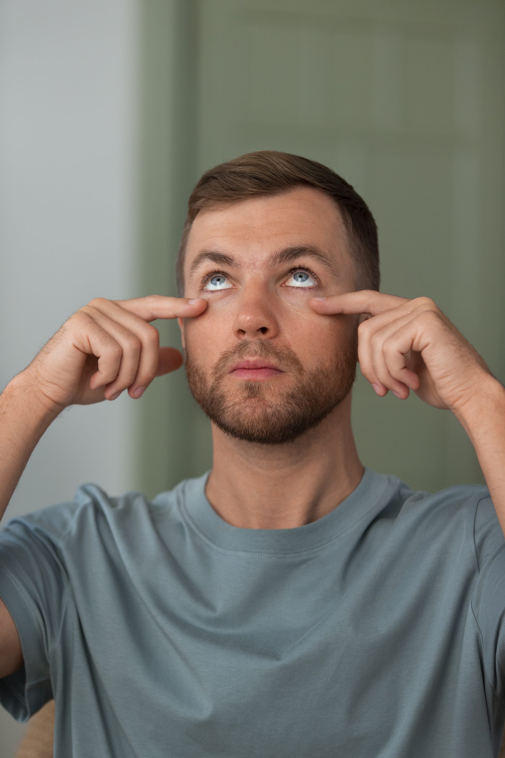
Function of the Orbicularis Oculi
Close your eyes and feel the orbicularis oculi muscle effortlessly bring your eyelids together. The orbicularis oculi muscle plays a crucial role in the closure of the eyelids, ensuring the protection and lubrication of the eyes. Its importance in eyelid closure cannot be overstated. Dysfunction of the orbicularis oculi muscle can occur due to various mechanisms and causes, leading to significant impacts on eye health. Disorders of the orbicularis oculi can result in conditions such as exposure keratitis and corneal problems if left untreated.
Fortunately, there are different treatment modalities available for orbicularis oculi muscle disorders. These include botulinum toxin injections, surgical interventions, physical therapy exercises, and medications. Advancements in surgical techniques have also contributed to improved outcomes for patients with orbicularis oculi muscle disorders.
Research in this field focuses on understanding the underlying mechanisms and causes of muscle dysfunction, as well as evaluating the efficacy of different treatment modalities. Collaborative efforts between researchers, clinicians, and patients are driving advancements in the field of orbicularis oculi muscle disorders. These advancements aim to provide targeted therapies and enhance the quality of life for individuals suffering from these conditions.
Embryological Development of the Orbicularis Oculi
As you explore the embryological development of the orbicularis oculi, it becomes evident that this essential eye muscle originates from the mesoderm in the eyelids during the twelfth week of development. The development of the orbicularis oculi involves several key processes and factors, including:
- Developmental timeline:
- The orbicularis oculi muscle begins to form during the twelfth week of embryological development.
- By the fifteenth week, the muscle is well-developed and functional.
- Mesoderm origin:
- The orbicularis oculi muscle arises from the mesoderm, which is one of the three primary germ layers during embryogenesis.
- Specifically, it originates from the mesenchyme of the second pharyngeal arch.
- Neural crest involvement:
- The neural crest cells, a group of multipotent cells derived from the embryonic ectoderm, play a role in the development of the orbicularis oculi muscle.
- These cells migrate and contribute to the formation of the facial muscles, including the orbicularis oculi.
Other important aspects of the embryological development of the orbicularis oculi include the vascularization process and the influence of genetic factors. The muscle receives its blood supply from branches of the facial artery, superficial temporal artery, and ophthalmic artery. Additionally, genetic factors play a crucial role in the proper development and functioning of the orbicularis oculi muscle. Understanding the embryological development of this muscle is essential for comprehending its role in eyelid closure and its potential involvement in various eye-related disorders.
Blood Supply to the Orbicularis Oculi
Exploring the blood supply to the orbicularis oculi muscle reveals its essential role in nourishing and supporting the function of this crucial eye muscle. The blood vessels that supply the orbicularis oculi play a significant role in maintaining the muscle’s function and overall health. The facial artery, superficial temporal artery, and ophthalmic artery are responsible for providing the necessary blood supply to this muscle.
The blood vessels deliver oxygen and nutrients to the orbicularis oculi, ensuring its proper function in tear drainage and eye closure. Additionally, these vessels also remove waste products and toxins from the muscle, promoting its optimal performance.
The involvement of the facial nerve in the innervation of the orbicularis oculi further emphasizes the importance of blood supply in supporting the muscle’s function. The facial nerve branches, such as the temporal and zygomatic branches, provide the necessary signals for the muscle to contract and facilitate eyelid closure.
In cases of orbicularis oculi disorders, surgical interventions may be necessary to address any issues related to blood supply. These interventions can help restore proper circulation to the muscle, ensuring its ability to perform its role in tear drainage and eye closure effectively.
Innervation of the Orbicularis Oculi
To understand the function and role of the orbicularis oculi muscle, it is important to explore its innervation and how it allows for proper eyelid closure. The innervation of the orbicularis oculi muscle is provided by the facial nerve, specifically the zygomatic and temporal branches. Here is a breakdown of the innervation process:
- Facial nerve: The orbicularis oculi muscle receives innervation from the facial nerve, which is the seventh cranial nerve. This nerve plays a crucial role in controlling the muscles of facial expression.
- Zygomatic branch: The upper half of the orbicularis oculi muscle is innervated by the zygomatic branch of the facial nerve. This branch controls the voluntary closure of the upper eyelid.
- Temporal branch: The lower half of the orbicularis oculi muscle is innervated by the temporal branch of the facial nerve. This branch controls the voluntary closure of the lower eyelid.
Proper innervation of the orbicularis oculi muscle is essential for eyelid closure and the normal functioning of the blinking mechanism. Facial paralysis or damage to the facial nerve can lead to difficulty in closing the eyelids, resulting in incomplete eyelid closure and potential eye problems. Understanding the muscle’s innervation helps in diagnosing and treating conditions related to the orbicularis oculi muscle and ensuring proper eyelid closure.
Muscles and Subdivisions of the Orbicularis Oculi
The orbicularis oculi muscle consists of various muscles and subdivisions that play a crucial role in the eyelid closure and overall functioning of the eye. This muscle is responsible for closing the eyelids, which is essential for protecting the eye from external irritants and maintaining tear film distribution. The orbicularis oculi muscle is innervated by the facial nerve, specifically the zygomatic and temporal branches.
Surgical interventions may be necessary in cases of dysfunctional orbicularis oculi muscle, such as in the treatment of conditions like Bell’s palsy or facial nerve injuries. These interventions aim to restore proper eyelid closure and prevent complications such as corneal problems and exposure keratitis.
Corneal problems can arise when the eyelids fail to close properly, leading to exposure of the cornea and increased risk of corneal ulceration, scarring, and perforation. It is important to address orbicularis oculi muscle dysfunction promptly to prevent these complications.
Clinical Significance of the Orbicularis Oculi
When assessing the clinical significance of the orbicularis oculi muscle, it is important to consider its role in eyelid closure and its potential impact on corneal health. The muscle plays a crucial role in maintaining the integrity of the eyelids and ensuring proper closure. Here are some key points regarding the clinical significance of the orbicularis oculi:
- Impact of facial nerve paralysis on orbicularis oculi function: Facial nerve paralysis can result in weakness or complete loss of function of the orbicularis oculi muscle, leading to difficulties in eyelid closure. This can cause exposure of the cornea, increasing the risk of corneal damage and associated complications.
- Role of the orbicularis oculi in tear drainage and eye health: The orbicularis oculi muscle assists in tear drainage by compressing the lacrimal sac during blinking. Dysfunction of this muscle can impair tear drainage, leading to excessive tearing or dry eye syndrome.
- Evaluation and management of blepharospasm associated with orbicularis oculi dysfunction: Blepharospasm is a condition characterized by involuntary contractions of the orbicularis oculi muscle, resulting in uncontrollable eyelid blinking or closure. Treatment options include botulinum toxin injections, medications, and surgical interventions.
- Age-related changes in the orbicularis oculi muscle and its effects on eyelid closure: With aging, the orbicularis oculi muscle may undergo changes, such as muscle laxity or weakening. These changes can affect eyelid closure and contribute to age-related eyelid abnormalities, such as drooping or sagging.
- Comparison of different treatment modalities for orbicularis oculi muscle disorders: Treatment options for orbicularis oculi muscle disorders include botulinum toxin injections, surgical interventions, physical therapy exercises, and medications. The choice of treatment depends on the specific condition, severity of symptoms, and individual patient factors.
Considering the clinical significance of the orbicularis oculi muscle is crucial in the evaluation, management, and treatment of various conditions affecting eyelid closure and eye health. Understanding its role and potential dysfunctions allows for targeted interventions to improve patient outcomes.
Treatment Options for Orbicularis Oculi Disorders
If you are experiencing disorders related to the orbicularis oculi muscle, there are various treatment options available to address your specific condition and improve your overall eye health. One common treatment option is the use of botulinum toxin injections, which have shown efficacy in treating orbicularis oculi disorders. These injections work by temporarily paralyzing the muscle and reducing its activity, which can help alleviate symptoms such as eyelid spasms or excessive blinking. Another treatment option for orbicularis oculi muscle dysfunction is surgical intervention. In cases where the muscle is severely impaired or damaged, surgical procedures may be necessary to repair or reconstruct the muscle. Physical therapy exercises can also be beneficial for rehabilitating the orbicularis oculi muscle. These exercises focus on strengthening the muscle and improving its coordination and control. Additionally, medications may be prescribed to manage orbicularis oculi muscle disorders. These medications can help reduce inflammation, relieve pain, or address underlying conditions that may be contributing to the muscle dysfunction. Finally, advancements in targeted therapies are being explored for the treatment of orbicularis oculi muscle dysfunction. These therapies aim to specifically target and correct the underlying causes of the muscle disorder, offering potential long-term solutions for patients. It is important to consult with a healthcare professional to determine the most appropriate treatment option for your specific condition.
Complications Associated With Orbicularis Oculi Disorders
What are the potential complications that can arise from orbicularis oculi disorders?
Complications associated with orbicularis oculi disorders can have significant impacts on both the function and appearance of the eyes. Here are some potential complications to be aware of:
- Corneal problems: Poor closure of the eyelids can lead to exposure keratitis, which is inflammation of the cornea caused by insufficient protection and lubrication. This can result in corneal ulcers, scarring, and even corneal perforation if not treated adequately.
- Functional and cosmetic impairments: Disorders of the orbicularis oculi muscle can affect the ability to fully close the eyelids, leading to functional impairments such as difficulty in keeping the eyes moist and protecting them from external irritants. Additionally, cosmetic impairments may arise, including drooping of the eyelids or asymmetry in eyelid closure.
- Discomfort and pain: Patients with orbicularis oculi disorders may experience discomfort and pain in the eyes due to inadequate eyelid closure and exposure to environmental factors like wind, dust, and bright light.
Management and prevention of these complications involve a comprehensive approach. Current research focuses on understanding the underlying mechanisms and causes of muscle dysfunction, as well as advancements in surgical techniques and the development of targeted therapies. Patient experiences play a crucial role in improving treatment options and outcomes for orbicularis oculi disorders. By collaborating with researchers and clinicians, patients can contribute valuable insights that can lead to advancements in the field.
Research and Advancements in Orbicularis Oculi Disorders
To further explore the field of orbicularis oculi disorders, let’s delve into the latest research and advancements in understanding and treating these conditions. Research collaborations have played a crucial role in advancing our knowledge in this area. Scientists and clinicians are working together to investigate the underlying mechanisms and causes of orbicularis oculi muscle dysfunction. This collaborative effort has led to significant advancements in the field.
One of the most widely used treatments for orbicularis oculi disorders is botulinum toxin therapy. This therapy involves injecting small amounts of botulinum toxin into the muscle to temporarily relax it and reduce muscle spasms. It has shown promising results in improving eyelid closure and reducing symptoms.
Surgical advancements have also been made in the treatment of orbicularis oculi disorders. Surgeons have developed innovative techniques to repair and strengthen the muscle, improving eyelid function and appearance. These surgical interventions have provided effective solutions for patients with severe muscle dysfunction.
Physical therapy interventions have been explored as a non-invasive treatment option for orbicularis oculi disorders. Specific exercises and techniques can help improve muscle strength, coordination, and control, leading to improved eyelid closure.
In addition to these approaches, new medication developments are being explored to target the underlying causes of orbicularis oculi muscle dysfunction. Researchers are investigating potential drug therapies that can modulate muscle function and improve eyelid closure.
Overall, the field of orbicularis oculi disorders is continuously evolving, thanks to research collaborations, advancements in botulinum toxin therapy, surgical techniques, physical therapy interventions, and new medication developments. These advancements offer hope for individuals with orbicularis oculi muscle dysfunction and pave the way for improved treatment options in the future.
| Research Collaborations | Botulinum Toxin Therapy | Surgical Advancements | Physical Therapy Interventions |
|---|---|---|---|
| Ongoing studies focus on understanding the underlying mechanisms of muscle dysfunction. | Effective in reducing muscle spasms and improving eyelid closure. | Innovative techniques have improved muscle repair and strengthened the muscle. | Specific exercises and techniques improve muscle strength and control. |
| Collaboration between researchers and clinicians leads to advancements in the field. | Promising results in improving symptoms and function. | Surgical interventions provide solutions for severe muscle dysfunction. | Non-invasive treatment option for improving eyelid closure. |
| Investigating potential drug therapies to target the underlying causes of muscle dysfunction. | Ongoing research to optimize dosage and treatment protocols. | Improved techniques for better outcomes and patient satisfaction. | Enhances muscle coordination and control. |
| Important for sharing knowledge, resources, and expertise. | Widely used treatment option for orbicularis oculi disorders. | Provides options for patients who do not respond to other treatments. | Can be tailored to individual needs and goals. |
Table: Latest research and advancements in understanding and treating orbicularis oculi disorders.