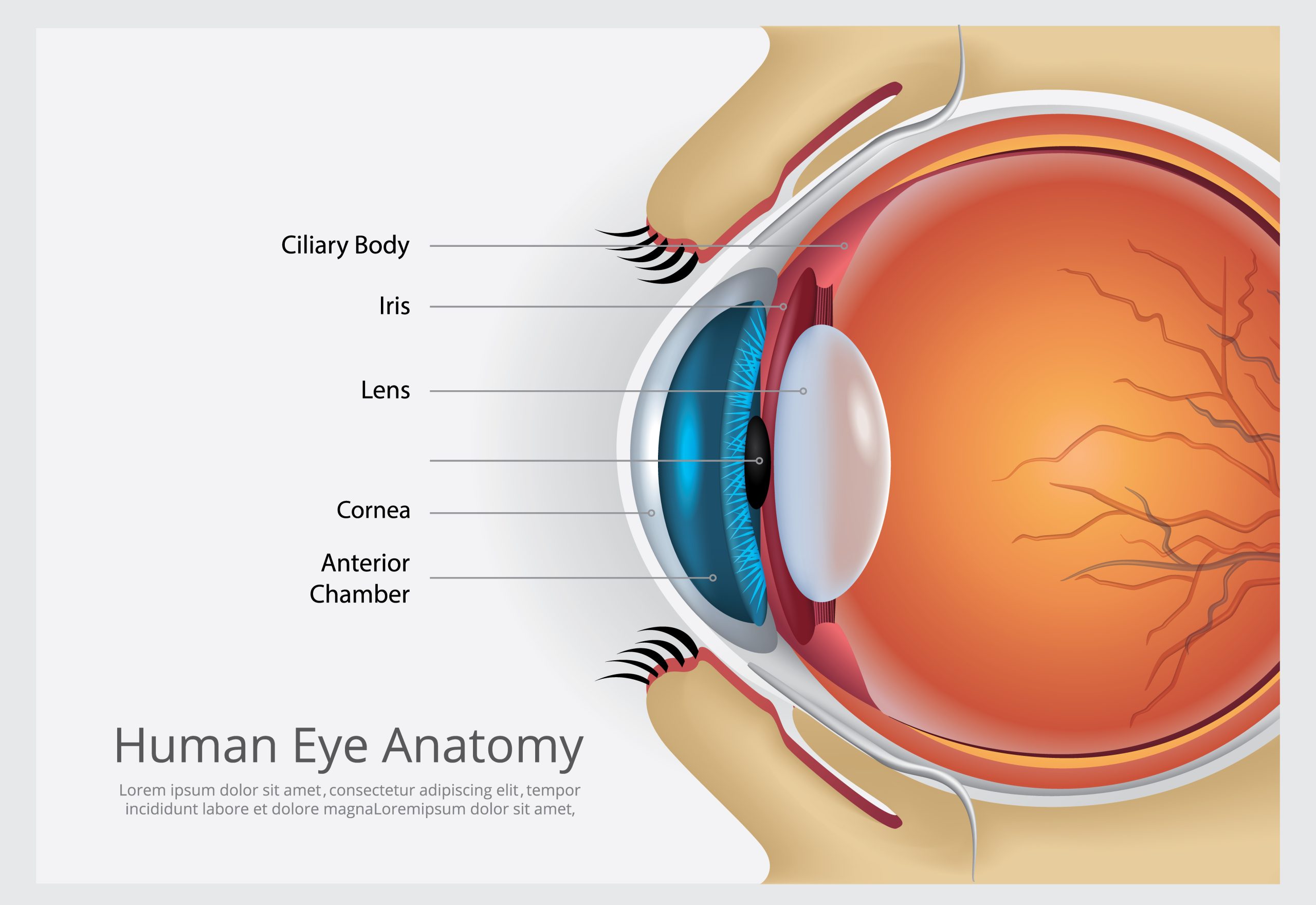
Have you ever wondered what makes your eyelids function so seamlessly? In this article, we’ll delve into the intricate details of the anatomy of the eyelid. From the layers of skin and subcutaneous tissue to the levator apparatus responsible for opening the lid, we’ll explore every component. We’ll also discuss the role of the conjunctiva, vasculature, and innervation in maintaining the health and functionality of the eyelid. By the end, you’ll have a comprehensive understanding of this essential part of your eye.
Layers of the Eyelid
Let’s now delve into the layers of the eyelid. The skin and subcutaneous tissue form the outermost layer, followed by the orbicularis oculi muscle responsible for eyelid closure. The tarsal plates provide structural support, while the levator apparatus, including the levator palpebrae superioris and superior tarsal muscles, open the eyelid. Lastly, the conjunctiva forms the deepest layer, offering protection and lubrication to the eye.
Skin and subcutaneous tissue
To understand the anatomy of the eyelid, it is important to examine the layers of the eyelid, specifically the skin and subcutaneous tissue. Here are some key points to consider:
- Eyelid thickness: The skin of the eyelid is the thinnest in the body, measuring less than 1 mm thick.
- Eyelid elasticity: The skin and subcutaneous tissue of the eyelid allow for easy distension, making it susceptible to eyelid edema.
- Eyelid edema: Swelling of the eyelid can occur due to various factors, such as allergies, infections, or trauma.
- Eyelid infections: Infections can affect the skin and subcutaneous tissue of the eyelid, leading to conditions like blepharitis or cellulitis.
- Eyelid trauma: Trauma to the eyelid can result in injuries such as lacerations or contusions, which may involve the skin and subcutaneous tissue.
Understanding the structure and characteristics of the skin and subcutaneous tissue of the eyelid is crucial in diagnosing and managing various eyelid conditions and injuries.
Orbicularis oculi
Eyelid thickness plays a crucial role in the function and structure of the orbicularis oculi muscle. The orbicularis oculi is the main muscle responsible for eyelid closure and is involved in various facial expressions. It consists of three parts: the palpebral, lacrimal, and orbital. The palpebral part gently closes the eyelids, while the lacrimal part is involved in tear drainage, and the orbital part tightly closes the eyelids. The orbicularis oculi muscle is innervated by the facial nerve and has a close relationship with other facial muscles.
To provide a deeper understanding, here is a table summarizing the key aspects of the orbicularis oculi muscle:
| Aspect | Description |
|---|---|
| Function | Responsible for eyelid closure and various facial expressions |
| Innervation | Facial nerve |
| Parts | Palpebral, lacrimal, and orbital |
| Palpebral Part | Gently closes the eyelids |
| Lacrimal Part | Involved in tear drainage |
| Orbital Part | Tightly closes the eyelids |
The orbicularis oculi muscle is an essential component of the eyelid, contributing to its function and allowing for proper eyelid closure and a wide range of facial expressions.
Tarsal plates
The tarsal plates, located deep within the eyelids, serve as the structural framework for the eyelid. These plates are composed of dense connective tissue and have a crescentic shape. They play a crucial role in the function and anatomy of the eyelids. Here are some key points about tarsal plates:
- Tarsal plate function: The tarsal plates provide support and stability to the eyelids, allowing them to open and close smoothly. They also serve as the attachment site for muscles that control eyelid movement.
- Tarsal plate structure: Each eyelid has two tarsal plates – the superior tarsus in the upper eyelid and the inferior tarsus in the lower eyelid. They are attached to the margin of the orbits through medial and lateral palpebral ligaments.
- Tarsal plate disorders: Disorders of the tarsal plates, such as tarsal plate laxity or entropion, can lead to eyelid malposition and functional problems.
- Tarsal plate development: The tarsal plates develop during embryogenesis and undergo maturation throughout childhood and adolescence.
- Tarsal plate anatomy: The tarsal plates contain Meibomian glands, which secrete oils that help prevent tear evaporation and keep the eyelids lubricated.
Understanding the structure and function of the tarsal plates is essential for comprehending the complex anatomy of the eyelids.
Levator apparatus
Moving on to the next layer of the eyelid, let’s delve into the fascinating world of the levator apparatus. The levator apparatus consists of the levator palpebrae superioris and the superior tarsal muscle, which play a crucial role in opening the eyelid. The levator palpebrae superioris originates from the lesser wing of the sphenoid bone and inserts into the upper eyelid and the superior tarsal plate. On the other hand, the superior tarsal muscle originates from the underside of the levator palpebrae superioris and inserts into the superior tarsal plate. These muscles are innervated by the superior branch of the oculomotor nerve (CN III) and sympathetic fibers from the superior cervical ganglion. Together, these muscles work in coordination to elevate the eyelid.
Conjunctiva
Delving deeper into the layers of the eyelid, let’s now explore the role of the conjunctiva in providing protection and lubrication to the eye.
- Conjunctiva: Functions:
- The conjunctiva is a thin mucous membrane that lines the inner surface of the eyelid (palpebral conjunctiva) and covers the sclera of the eyeball (bulbar conjunctiva).
- It provides protection to the eye by acting as a barrier against foreign particles and pathogens.
- The conjunctiva also helps in lubricating the eye by secreting mucus and tears.
- It plays a crucial role in maintaining the health and integrity of the ocular surface.
- Conjunctiva: Anatomy and Physiology:
- The conjunctiva is a thin, transparent membrane composed of epithelial cells and underlying connective tissue.
- It contains numerous blood vessels that supply oxygen and nutrients to the ocular surface.
- The conjunctiva also houses specialized cells called goblet cells, which produce mucus to keep the eye moist and protected.
- Conjunctiva: Clinical Examination:
- During a clinical examination, the conjunctiva is carefully examined for any signs of inflammation, infection, or tumors.
- The color, texture, and vascular pattern of the conjunctiva are assessed to evaluate the overall health of the eye.
- Conjunctiva: Inflammatory Conditions:
- Conjunctivitis, or inflammation of the conjunctiva, is a common condition that can be caused by bacterial or viral infections, allergies, or irritants.
- Other inflammatory conditions of the conjunctiva include conjunctival cysts, pinguecula, and pterygium.
- Conjunctiva: Surgical Procedures:
- Surgical procedures involving the conjunctiva may be performed to treat certain conditions, such as pterygium or conjunctival tumors.
- These procedures may include conjunctival grafting, excision of abnormal tissue, or reconstruction of the conjunctiva.
Understanding the anatomy and function of the conjunctiva is essential for the evaluation and management of various ocular conditions. From its role in protection and lubrication to its involvement in inflammatory conditions and surgical procedures, the conjunctiva plays a vital role in maintaining the health of the eye.
Levator Apparatus
Elevate your understanding of the eyelid’s anatomy by exploring the function of the levator apparatus. The levator palpebrae superioris and superior tarsal muscles play a crucial role in opening the upper eyelid. The levator palpebrae superioris originates from the lesser wing of the sphenoid bone and inserts into the upper eyelid and the superior tarsal plate. On the other hand, the superior tarsal muscle originates from the underside of the levator palpebrae superioris and inserts into the superior tarsal plate. These muscles are innervated by the superior branch of the oculomotor nerve (CN III) and sympathetic fibers from the superior cervical ganglion.
Understanding the levator apparatus is essential in various aspects of ophthalmology. Levator dysfunction, for example, can lead to ptosis or drooping of the upper eyelid. Levator surgery, such as levator resection or levator advancement, is often performed to correct ptosis and improve eyelid function. In-depth knowledge of the levator anatomy and innervation is necessary for accurate surgical planning and execution. By grasping the intricacies of the levator apparatus, you can gain a comprehensive understanding of the eyelid’s anatomy and its impact on ocular health and function.
Conjunctiva
Now let’s explore the role of the conjunctiva, a thin mucous membrane, in protecting and lubricating the eye.
- The conjunctiva has several functions, including protecting the eye from foreign particles and infection, lubricating the surface of the eye, and aiding in tear production.
- It is located on the inner surface of the eyelids (palpebral conjunctiva) and extends onto the white part of the eye (bulbar conjunctiva).
- The conjunctiva has a close relationship with the sclera, the tough outer layer of the eye. It covers and protects the sclera, providing a smooth surface for the eyelids to move over.
- The conjunctiva plays a crucial role in tear production. It contains goblet cells that secrete mucus, which helps to spread tears evenly over the surface of the eye.
- Diseases of the conjunctiva can include conjunctivitis (inflammation of the conjunctiva), pterygium (growth on the conjunctiva), and pinguecula (yellowish deposit on the conjunctiva).
The conjunctiva is an important structure in the eye, contributing to its overall health and function.
Vasculature
To understand the vasculature of the eyelid, let’s explore its rich arterial supply and venous drainage. The eyelid receives a robust vascular supply from various arteries. The ophthalmic artery is the primary supplier, providing arterial branches such as the lacrimal, medial palpebral, supraorbital, dorsal nasal, and supratrochlear arteries. Additionally, the facial artery contributes through its angular branch, while the superficial temporal artery supplies the transverse facial artery branch.
Venous drainage of the eyelid occurs through several veins, including the medial palpebral vein, angular vein, ophthalmic vein, lateral palpebral vein, and superficial temporal vein.
In terms of the lymphatic system, the upper eyelid and outer half of the lower eyelid drain into pre-auricular lymph nodes, while the middle of the upper eyelid and inner half of the lower eyelid drain into submandibular lymph nodes.
Furthermore, the lacrimal drainage system plays a vital role in tear drainage. Tears are produced by the lacrimal gland and drain through the lacrimal puncta, canaliculi, lacrimal sac, and nasolacrimal duct. Dysfunction of the lacrimal glands can result in dry eyes or excessive tearing.
Understanding the vasculature of the eyelid is crucial for comprehending its physiological processes and potential pathological conditions related to vascular supply, arterial branches, venous drainage, lymphatic system, and lacrimal drainage.
Innervation
Moving on to the innervation of the eyelid, let’s delve into the sensory and motor components that provide functionality to this intricate anatomical structure.
- Sensory innervation to the eyelids is provided by branches of the trigeminal nerve. The upper eyelid is supplied by the ophthalmic nerve (V1), including the supraorbital, supratrochlear, infratrochlear, and lacrimal branches. The lower eyelid is supplied by the maxillary nerve (V2), including the infraorbital and zygomaticofacial branches.
- Motor innervation to the muscles of the eyelid is provided by the facial nerve (orbicularis oculi), oculomotor nerve (levator palpebrae superioris), and sympathetic fibers (superior tarsal muscle).
- The lacrimal gland, responsible for tear production, is supplied by the lacrimal nerve, a branch of the ophthalmic division of the trigeminal nerve.
- Sensory innervation allows for the perception of touch, pain, and temperature in the eyelid, providing protective reflexes and awareness of the environment.
- Motor innervation controls the movement of the eyelid muscles, allowing for blinking, closure, and opening, as well as the regulation of tear production through the lacrimal system.
Lacrimal Glands
The lacrimal glands, responsible for tear production, play a crucial role in maintaining the functionality of the eyelid. These glands are located in the superolateral aspect of the orbit and are responsible for producing tears, which lubricate the eye and help wash away foreign particles. Tears are composed of water, electrolytes, proteins, and lipids, and their production is regulated by the lacrimal system.
The lacrimal system consists of the lacrimal glands, tear drainage pathways, the lacrimal sac, and the nasolacrimal duct. Tears are produced by the lacrimal glands and are spread across the surface of the eye with each blink. Excess tears are drained through the lacrimal puncta, into the canaliculi, then into the lacrimal sac, and finally into the nasolacrimal duct, which empties into the nasal cavity.
The function of the lacrimal glands is essential for maintaining the health of the eye. Tear production helps to keep the surface of the eye moist, preventing dryness and irritation. Additionally, tears contain antibodies and enzymes that protect the eye from infections. Dysfunction of the lacrimal glands can lead to dry eyes or excessive tearing, both of which can impact vision and overall eye health. Therefore, the proper functioning of the lacrimal glands is crucial for maintaining the integrity and functionality of the eyelid.
Eyelid and Orbit Anatomy
As we delve into the topic of eyelid and orbit anatomy, let’s continue our exploration of the intricate structures that contribute to the functionality and protection of the eyelid by examining their composition and organization.
- Eyelid function: The eyelids serve multiple purposes, including protection, light control, and lubrication of the eye.
- Eyelid structure: The eyelids consist of skin, subcutaneous tissue, orbicularis oculi muscle, septum, tarsi, and fat tissue.
- Eyelid development: The development of the eyelids involves the fusion of the upper and lower eyelid folds during embryogenesis.
- Eyelid disorders: Various disorders can affect the eyelids, such as ptosis (drooping eyelids), ectropion (outward turning of the eyelid), and entropion (inward turning of the eyelid).
- Eyelid surgery: Surgical procedures can be performed on the eyelids to correct functional or cosmetic issues, such as blepharoplasty (eyelid lift) or ptosis repair.
Understanding the anatomy of the eyelids and orbit is crucial for diagnosing and treating eyelid disorders and performing eyelid surgery effectively. By studying the intricate structures and their organization, healthcare professionals can ensure optimal outcomes for their patients.
Tarsal Plates
Continue exploring the anatomy of the eyelid by delving into the composition and function of the tarsal plates. Tarsal plates are dense fibrous tissues located deep to the palpebral region of the orbicularis oculi muscle. They serve as the scaffolding of the eyelid and are composed of dense connective tissue. The eyelid has two tarsal plates: the superior tarsus in the upper eyelid and the inferior tarsus in the lower eyelid.
To provide a clearer understanding, here is a table summarizing the key aspects of tarsal plates:
| Tarsal Plates | Function |
|---|---|
| Structure | Dense connective tissue forming the scaffolding of the eyelid |
| Clinical Significance | Provide support and stability to the eyelid; serve as the attachment site for muscles and glands |
| Development and Growth | Tarsal plates develop and grow along with the eyelid during embryonic and postnatal stages |
| Aging and Changes | Tarsal plates undergo changes with age, such as loss of elasticity and thinning of tissues |
| Surgical Procedures and Interventions | Tarsal plates are involved in various eyelid surgeries, such as ptosis repair and blepharoplasty |
Understanding the function, structure, clinical significance, development, aging, and surgical relevance of tarsal plates is crucial in comprehending the intricate anatomy of the eyelid.
Orbital Septum
To understand the anatomy of the eyelid in more detail, let’s explore the role and composition of the orbital septum. The orbital septum is a connective tissue band that separates the eyelids from the orbit. It is attached to the border of the orbital bone and lid retractors. The orbital septum contains multiple layers in close relationship with the anterior connective tissue framework and extends along the rim of the orbit and supraorbital groove. It also extends to the anterior lacrimal crest and lateral orbital margin.
The orbital septum plays a crucial role in eyelid function. Here are the key functions and features associated with the orbital septum:
- Separates the eyelids from the orbit, providing structural support and maintaining the shape of the eyelids.
- Acts as a barrier between the orbital contents and the eyelids, protecting the delicate structures within the orbit.
- Provides a attachment site for the lacrimal ducts, which drain tears from the eyes.
- Contributes to eyelid thickness, playing a role in the overall appearance and contour of the eyelids.
- Works in conjunction with the tarsal plates to maintain the integrity and stability of the eyelids.
Understanding the role and composition of the orbital septum is essential for comprehending the intricate anatomy of the eyelid and its many functions.
Blood Supply, Lymphatic Drainage, Muscles, and Bones
Now, let’s delve into the intricate details of the blood supply, lymphatic drainage, muscles, and bones that contribute to the complex anatomy of the eyelid. The eyelid vasculature is supplied by branches of the internal and external carotid arteries. The ophthalmic artery, a branch of the internal carotid artery, provides blood supply to different parts of the eyelid. The superior and inferior marginal vessels form the marginal arcade, located at the front of the tarsus. The lacrimal artery passes through the orbital septum and joins the marginal arcade. Lymphatic drainage of the eyelid occurs in two different pathways. The upper eyelid and outer half of the lower eyelid drain into pre-auricular lymph nodes, while the middle of the upper eyelid and inner half of the lower eyelid drain into submandibular lymph nodes. The muscles responsible for eyelid movement include the orbicularis oculi, levator palpebrae superioris, and the extraocular muscles. The orbicularis oculi muscle closes the eyelids, while the levator palpebrae superioris elevates the upper eyelid. The extraocular muscles control different movements of the eyeball and are innervated by the oculomotor, trochlear, and abducens nerves. The eyelid and orbit anatomy is also influenced by the presence of the lacrimal system, which includes the lacrimal gland responsible for tear production. The lacrimal gland is supplied by the lacrimal nerve, a branch of the ophthalmic division of the trigeminal nerve.


