Well, get ready to uncover the secrets behind this condition. In this discussion, we will delve into the four types of entropion: congenital, involutional, acute spastic, and cicatricial. But that’s not all – we’ll also explore the underlying causes, evaluation methods, and treatment options. Medical curiosity, especially about conditions that affect you, is one of the more intellectual ways to engage in personal discovery and self-reflection. So, if you’re ready to gain a deeper understanding of entropion and its implications, let’s dive right in.
Congenital Entropion
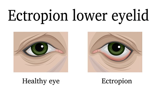
Congenital entropion is characterized by the inward turning of the eyelid margin from birth, often causing discomfort and potential corneal and conjunctival damage. It is a rare condition that can be caused by various factors. The exact cause of congenital entropion is not fully understood, but it is believed to be due to abnormal development of the eyelid structures during fetal development. This can result in a misalignment of the eyelid margin, leading to the inward turning.
Symptoms of congenital entropion may include excessive tearing, redness, and irritation of the eye. The condition can also lead to corneal abrasions, infections, and scarring if left untreated.
Surgical options are available for the treatment of congenital entropion. The surgical procedure aims to correct the misalignment of the eyelid margin and restore its normal position. This can be achieved through techniques such as eyelid repositioning, eyelid tightening, or grafting.
Complications of congenital entropion surgery may include infection, bleeding, scarring, or recurrence of the condition. However, with proper preoperative evaluation and postoperative care, the risk of complications can be minimized.
Involutional Entropion
Involutional entropion is a common eyelid malposition characterized by the inward turning of the eyelid margin, predominantly seen in the elderly population. This condition can cause significant discomfort and visual disturbances. Here are some important points to understand about involutional entropion:
Causes:
- Involutional changes associated with aging, such as horizontal laxity of the eyelid, weakening or disinsertion of eyelid retractors, and overriding by the preseptal orbicularis oculi muscle, contribute to the development of involutional entropion.
Symptoms:
- Patients with involutional entropion often experience foreign body sensation, redness, tearing, and discharge. Dry eye syndrome and corneal abrasions can also occur.
Surgical Options:
- Surgical correction is the main treatment option for involutional entropion. It involves exploration and repair of the lower eyelid retractors, resection of the pretarsal orbicularis oculi muscle, and canthal tightening procedures.
Complications:
- If left untreated, involutional entropion can lead to corneal and conjunctival damage, including corneal stromal abrasion, scarring, thinning, neovascularization, and even ulceration or perforation.
Prevention:
- There are no specific preventive measures for involutional entropion. However, early detection and timely surgical intervention can help prevent complications and improve quality of life for affected individuals.
Acute Spastic Entropion
Acute spastic entropion, unlike involutional entropion, is characterized by a temporary inward turning of the eyelid margin caused by sustained orbicularis oculi muscle contraction. It is often caused by ocular irritation or inflammation. The main symptom of acute spastic entropion is a foreign body sensation in the eye, along with redness, tearing, and discharge. The treatment options for acute spastic entropion include medical management and surgical intervention. In mild cases, ocular lubrication and artificial tears can provide relief. Botulinum toxin injection can also be used to temporarily paralyze the orbicularis muscle and alleviate the muscle contraction. In more severe cases, surgical correction may be necessary. Complications of acute spastic entropion include corneal and conjunctival damage, which can lead to corneal stromal abrasion, scarring, thinning, neovascularization, ulcer, and perforation. Prevention measures for acute spastic entropion include avoiding or managing conditions that can cause ocular irritation or inflammation, such as dry eye syndrome or infections. It is important to consult with a healthcare professional for a proper diagnosis and appropriate treatment plan for acute spastic entropion.
Cicatricial Entropion
Cicatricial entropion is characterized by the inward turning of the eyelid margin, which is caused by tarsal conjunctival contracture and internal rotation of the eyelid margin. This condition can lead to various complications if left untreated. Here are some key points to help you understand cicatricial entropion:
- Causes: Cicatricial entropion is often caused by tarsal conjunctival contracture, which results in the shortening and tightening of the tissue. This can occur due to scarring from previous surgeries, trauma, or inflammation.
- Complications: If left untreated, cicatricial entropion can cause corneal and conjunctival damage. This can lead to corneal stromal abrasion, scarring, thinning, neovascularization, ulcer, and even perforation.
- Surgical options: Surgical correction is often necessary for cicatricial entropion. The specific procedure will depend on the severity of the condition and the underlying cause. Options may include exploration and repair of the lower eyelid retractors, resection of pretarsal orbicularis oculi, and canthal tightening procedures.
- Non-surgical treatments: Before considering surgical correction, medical management of the underlying cause should be undertaken. This may include the use of ocular lubrication, artificial tears, or contact lenses.
- Prevention measures: Preventing cicatricial entropion involves minimizing the risk factors that can lead to tarsal conjunctival contracture. This includes proper wound care after surgeries, prompt treatment of infections and inflammation, and avoiding trauma to the eyelids.
Etiology and Pathophysiology
Entropion, the inward turning of the eyelid margin, can be caused by various factors that lead to the inversion of the lid margin and subsequent complications. These factors include horizontal eyelid laxity, attenuation of eyelid retractors, overriding by the preseptal orbicularis oculi muscle, previous surgeries and infections, and congenital origins.
Horizontal eyelid laxity refers to the looseness of the eyelid structures, such as the canthal tendons and tarsal plates. When these structures become lax, the lid margin is more prone to rotate inward, resulting in entropion. Attenuation of eyelid retractors, which provide vertical stability to the lower lid, can also contribute to entropion. When these retractors weaken, the lid is not properly supported, leading to the inward turning of the margin.
The preseptal orbicularis oculi muscle, which surrounds the eyelid, can override the eyelid margin and cause it to invert. This muscle contraction can be a contributing factor to spastic entropion.
Previous surgeries and infections can cause scarring and tissue damage, leading to entropion. Congenital origins, such as developmental abnormalities, can also result in the inward turning of the eyelid margin.
Understanding the etiology and pathophysiology of entropion is crucial for proper diagnosis and management of the condition. By addressing the underlying factors contributing to entropion, healthcare professionals can develop effective treatment plans to alleviate symptoms and prevent complications.
Epidemiology and Evaluation
The epidemiology and evaluation of entropion involves understanding its prevalence and conducting a comprehensive examination to determine the underlying causes and extent of the condition.
- Prevalence:
- Entropion is more common in the elderly population, with a higher prevalence in individuals over the age of 60.
- The prevalence increases with age, with 0.9% for individuals aged 60-69, 2.1% for those aged 70-79, and 7.6% for individuals over 80.
- Bilateral entropion is three times more common than unilateral.
- Women have a higher prevalence of entropion, with 2.4% compared to 1.9% in men.
- Involutional entropion has a reported prevalence of 2.4% in whites and 0.8% in blacks.
- Risk Factors:
- Age is a significant risk factor for developing entropion, as the condition is more prevalent in the elderly population.
- Women have a higher risk of developing entropion compared to men, possibly due to differences in tarsal plate size.
- Other risk factors include canthal tendon laxity, lower lid retractor issues, tarsal atrophy, and enophthalmos.
To evaluate entropion, a comprehensive examination is necessary. This includes assessing symptoms such as foreign body sensation, redness, tearing, and discharge. Physical examination involves evaluating for associated conditions like trichiasis, distichiasis, and epiblepharon. Special tests like corneal examination with fluorescein can help identify corneal damage, while conjunctival examination assesses for inflammation and margin abnormalities. The evaluation aims to determine the underlying cause and severity of entropion, which guides the appropriate management and treatment options.
Treatment and Management
To effectively manage entropion, the next step is to explore the treatment options available based on the underlying causes and severity of the condition. There are various approaches to treating entropion, including surgical interventions and medical management. Surgical correction is often necessary for severe or persistent cases, while medical management can provide temporary relief or be used as a first-line treatment in certain situations.
One option for medical management is the use of botulinum toxin injection, which can temporarily paralyze the orbicularis muscle and alleviate symptoms in cases of spastic entropion. Ocular lubrication, artificial tears, or contact lenses may also be recommended to improve symptoms and prevent further damage to the cornea and conjunctiva.
In cases of cicatricial entropion, medical management of the underlying cause should be undertaken before considering surgical correction. It is important to identify and address any infections or autoimmune diseases that may be contributing to the condition.
Surgical interventions for entropion may involve exploration and repair of the lower eyelid retractors, resection of the pretarsal orbicularis oculi, and canthal tightening procedures. In some cases of upper lid cicatricial entropion, tarsus replacement using grafts like tarsoconjunctival, scleral, or mucosal grafts may be necessary.
Interprofessional team strategies involving collaboration between healthcare professionals, such as ophthalmologists, facial plastics surgeons, and nurses, can lead to better outcomes in entropion management. Differential diagnosis is crucial to ensure that entropion is distinguished from other similar conditions, such as epiblepharon, trichiasis, trachoma, and distichiasis.
To emphasize the treatment options available for entropion, the following table provides a summary of the different interventions:
| Treatment Options | Description |
|---|---|
| Surgical Interventions | Exploration and repair of lower eyelid retractors |
| Resection of pretarsal orbicularis oculi | |
| Canthal tightening procedures | |
| Medical Management | Ocular lubrication |
| Artificial tears | |
| Contact lenses | |
| Botulinum toxin injection |