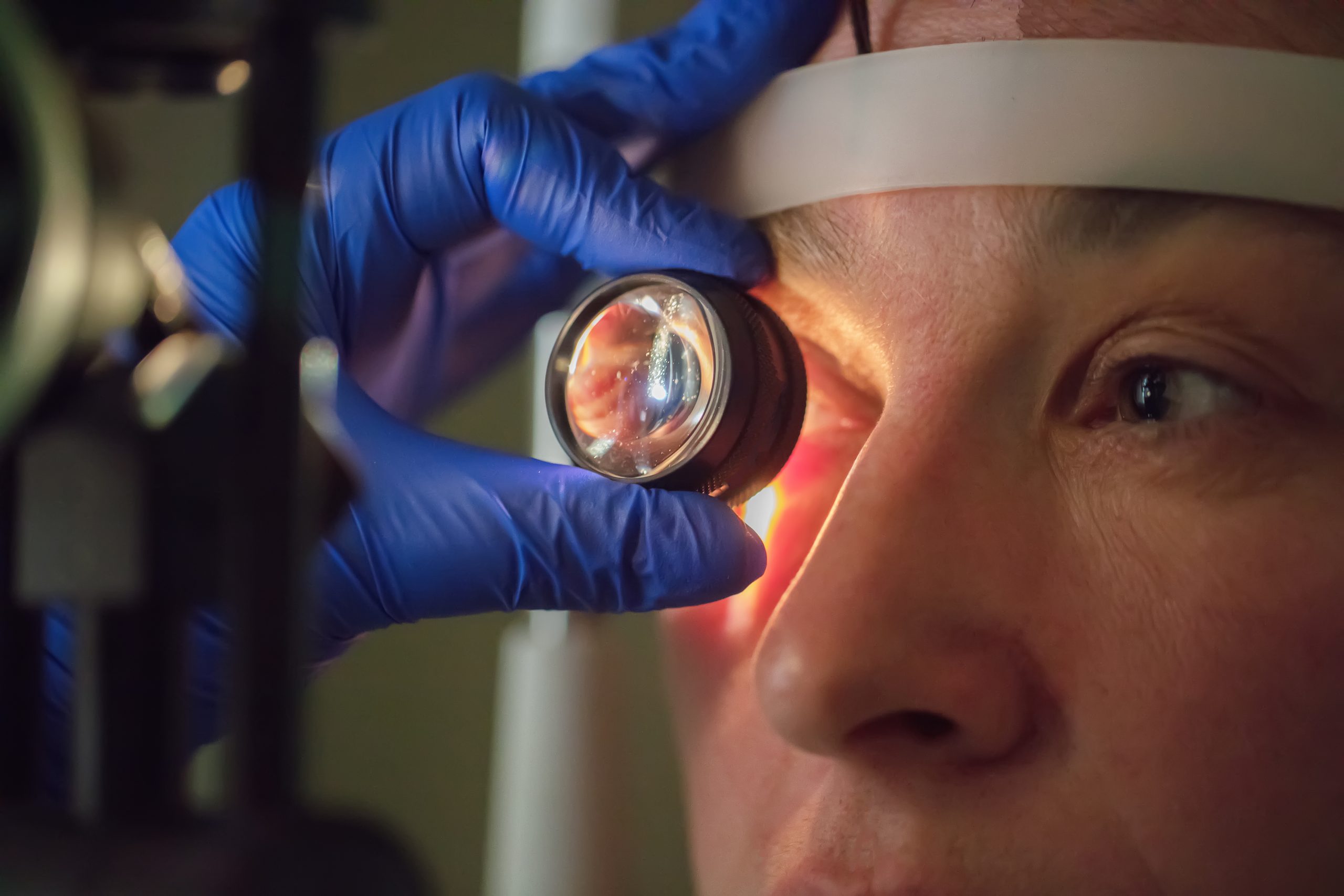
Are your eyes playing tricks on you? Blurred vision, haloes around lights, and that constant feeling of something in your eye could be signs of corneal edema. In this article, we’ll dive into the causes, symptoms, diagnosis, and treatment options for this condition. Corneal edema occurs when your cornea becomes overly hydrated, resulting in hazy vision and discomfort. It can be caused by damage to the endothelial cells, certain eye diseases, or surgical complications. To diagnose it, you’ll undergo visual acuity tests, slit-lamp examinations, and specialized imaging techniques. Treatment options range from simple saline drops to more invasive procedures like corneal transplantation. The healing time depends on the severity, but with regular follow-ups, you can find relief. Let’s explore recognizing the symptoms and the different treatment approaches for corneal edema.
Causes of Corneal Edema
Corneal edema is caused by various factors that lead to the swelling of the cornea. The most common causes of corneal edema include endothelial cell damage, Fuchs endothelial dystrophy, endotheliitis caused by the herpes virus, glaucoma, posterior polymorphous corneal dystrophy, Chandlers syndrome, cataract surgery, and use of certain drugs like benzalkonium chloride, chlorhexidine, and amantadine. Complications of corneal edema can include blurred or cloudy vision, hazy vision in the morning, haloes around lights, eye pain, feeling of a foreign object in the eye, eye redness, and sensitivity to light.
There are several risk factors for corneal edema, including older age, history of eye surgery, certain medical conditions like diabetes and uveitis, and prolonged use of contact lenses. It is important to note that corneal edema can be prevented by taking proper care of your eyes, avoiding eye injuries, and following your doctor’s instructions after eye surgery.
When it comes to treatment options for corneal edema, mild cases may not require treatment and can resolve on their own. However, in more severe cases, treatment options include temporary relief with concentrated saline drops or ointment, evaporating excess tears with a hair dryer (if safe), medications such as eye drops, hyperosmotic agents, corneal transplantation, Descemet’s stripping endothelial keratoplasty (DSEK), and endothelial keratoplasty (EK). The choice of treatment depends on the underlying cause and severity of the corneal edema.
Symptoms of Corneal Edema
If you’re experiencing corneal edema, you may notice symptoms such as sensitivity to light, seeing halos around lights, and the development of blisters on the cornea. These symptoms can greatly affect your vision and overall eye comfort. It’s important to seek medical attention if you’re experiencing any of these symptoms to determine the underlying cause and receive appropriate treatment.
Sensitivity to light
One common symptom of corneal edema is an increased sensitivity to light. This sensitivity, also known as photophobia, can cause discomfort and pain when exposed to bright lights or sunlight. Managing light sensitivity is an important aspect of corneal edema treatment. Wearing sunglasses or tinted lenses can help protect the eyes from excessive light and reduce discomfort. It is also crucial to prevent corneal edema complications by avoiding prolonged exposure to bright lights and taking breaks in dimly lit environments. Children with corneal edema may require special attention as their sensitivity to light can be more pronounced. Additionally, if you wear contact lenses, it is important to follow proper hygiene and care instructions to minimize the risk of corneal edema.
Seeing halos around lights
Are you experiencing halos around lights? Halos around lights are a common symptom of corneal edema. When the cornea becomes swollen and hydrated, it scatters light, causing the appearance of halos around light sources. There are several possible causes of halos, including endothelial cell damage, Fuchs endothelial dystrophy, glaucoma, and certain medications.
Managing halos involves treating the underlying cause of corneal edema. This may include using medications such as eye drops or hyperosmotic agents to reduce swelling. In severe cases, corneal transplantation or other surgical procedures may be necessary. Treatment for halos can help improve vision and reduce the impact on daily activities.
Preventing halos involves avoiding risk factors such as excessive contact lens wear, trauma to the eye, and certain medications. Following proper eye care practices, such as regular check-ups with an eye specialist, can also help prevent the development of corneal edema and halos. If you are experiencing halos around lights, it is important to seek medical attention for a proper diagnosis and treatment plan.
Blisters
Experiencing blisters on the cornea is a common symptom of corneal edema. These blisters, also known as subepithelial bullae, can be painful and cause discomfort. Here are some important points to know about blisters and corneal edema:
- Causes and prevention:
- Endothelial cell damage is a common cause of corneal edema and blisters.
- Other causes include Fuchs endothelial dystrophy, glaucoma, and cataract surgery.
- To prevent corneal edema and blisters, it is crucial to protect your eyes from injuries and avoid prolonged contact lens wear.
- Treatment options:
- Mild cases may not require treatment, but it’s important to manage symptoms.
- Medications, such as eye drops, can help relieve discomfort.
- In severe cases, corneal transplantation or other surgical procedures may be necessary.
- Long-term effects and managing symptoms:
- Corneal edema can lead to vision loss if left untreated.
- Regular follow-up with an eye specialist is important to monitor progress and manage symptoms effectively.
Remember to seek medical attention if you experience blisters or any other symptoms of corneal edema for proper diagnosis and treatment.
Diagnosing Corneal Edema
To diagnose corneal edema, your eye specialist will perform a series of tests and examinations. The purpose of these tests is to determine the cause of the corneal edema and assess its severity. The following table provides an overview of the diagnostic methods commonly used for corneal edema:
| Diagnostic Method | Description |
|---|---|
| Visual acuity test | Measures your ability to see clearly at different distances |
| Slit-lamp examination | Allows the doctor to examine the structures of your eye under magnification |
| Corneal pachymetry | Measures the thickness of the cornea |
| Specular microscopy | Provides detailed images of the corneal endothelial cells |
| Endothelial cell count | Determines the number and health of the endothelial cells |
These tests help the eye specialist diagnose corneal edema by evaluating the visual acuity, examining the cornea’s structure, and measuring its thickness and the health of the endothelial cells. Once the diagnosis is confirmed, appropriate treatment options can be discussed. Early diagnosis is crucial for effective management and prevention of complications. Regular follow-up with an eye specialist is important to monitor the progress and ensure the best possible outcome.
Treatment Options for Corneal Edema
You have several treatment options available for corneal edema. Here are three options to consider:
- Medications: Eye drops or ointments may be prescribed to manage corneal edema. These medications can help reduce swelling and improve vision. It’s important to follow the prescribed dosage and frequency.
- Hyperosmotic agents: These agents are used to draw fluid out of the cornea and reduce edema. They can be administered as eye drops or ointments. Hyperosmotic agents help relieve symptoms and improve vision in some cases.
- Corneal transplantation: In severe cases of corneal edema that do not respond to other treatments, a corneal transplantation may be necessary. This surgical procedure involves replacing the damaged cornea with a healthy donor cornea. It can help restore vision and alleviate symptoms.
It is important to consult with an eye specialist to determine the most suitable treatment approach for your specific case of corneal edema. They will consider factors such as the underlying cause, severity of the edema, and your overall eye health. Additionally, taking steps to prevent corneal edema, such as properly managing contact lenses and addressing any underlying eye conditions, can help reduce the risk of developing this condition and experiencing vision loss.
Healing Time and Recovery for Corneal Edema
The healing time and recovery for corneal edema depend on the severity of the condition and the chosen treatment approach. The length of time it takes for the cornea to heal and for vision to fully recover can vary. In mild cases of corneal edema, the condition may resolve within a few weeks. However, in severe cases, it may take several months for the cornea to heal completely.
The chosen treatment approach also plays a role in the healing time and recovery. Surgical options such as corneal transplantation, Descemet’s stripping endothelial keratoplasty (DSEK), and endothelial keratoplasty (EK) may require longer recovery periods compared to non-surgical treatments. Surgery to replace the entire cornea, for example, may take a year or longer for full vision recovery.
During the recovery process, it is important to monitor for any postoperative complications and adhere to any rehabilitation exercises recommended by your eye specialist. Regular follow-up appointments are crucial to assess progress and determine the long-term prognosis.
Outlook for Corneal Edema
After understanding the causes, symptoms, and treatment options for corneal edema, it is important to consider the outlook for this condition. The outlook for corneal edema depends on several factors, including the severity of the edema, the underlying cause, and the chosen treatment approach. Here are some key points to consider:
- Prognosis: Mild cases of corneal edema may progress slowly without causing noticeable symptoms. However, severe cases can lead to significant vision loss and complications.
- Long-term effects: If left untreated or poorly managed, corneal edema can result in permanent damage to the cornea and a decrease in vision quality. It can also increase the risk of developing other eye conditions.
- Preventive measures: While some risk factors for corneal edema, such as genetic conditions or certain eye surgeries, cannot be avoided, there are preventive measures that can help reduce the risk. These include proper eye hygiene, avoiding prolonged contact lens wear, and protecting the eyes from trauma or injury.
Corneal Edema: Definition and Effects
Corneal edema, characterized by swelling in the cornea, can have significant effects on vision and overall eye health. This condition occurs when the cornea becomes more hydrated than its normal 78% water content. Minor hydration changes may not impact the cornea significantly, but when the hydration level increases by 5% above normal, it can cause the cornea to scatter significant amounts of light and lose transparency. Corneal edema can also lead to a reduction in visual acuity and an increase in glare. The effects of corneal edema vary depending on the underlying cause, which can include endothelial cell damage, Fuchs endothelial dystrophy, glaucoma, and cataract surgery, among others. Treatment options for corneal edema range from mild cases not requiring treatment to medications, hyperosmotic agents, and even corneal transplantation in severe cases. It is important to manage corneal edema promptly to prevent complications and long-term effects on vision. Prevention strategies include avoiding factors that can contribute to corneal edema, such as trauma, hypoxia from contact lens wear, and high intraocular pressure. Regular follow-up with an eye specialist is essential for monitoring progress and adjusting treatment as needed.
Corneal Edema and Epithelial Dysfunction
To understand the relationship between corneal edema and epithelial dysfunction, it is important to recognize the impact of hydration changes on the cornea’s structure and function. Corneal edema, characterized by excess fluid accumulation in the cornea, can lead to various issues including hazy vision, increased glare, and reduction in visual acuity. Epithelial edema specifically affects visual acuity and can cause anterior irregular astigmatism. Here are some key points regarding corneal edema and epithelial dysfunction:
- Corneal Edema and Contact Lenses: Chronic hypoxia from overwearing contact lenses or poorly fitting lenses that alter corneal curvature can contribute to corneal edema. Improving tear exchange, redesigning the edge of the lens, or increasing lens size or optic zone can help alleviate the problem.
- Corneal Edema in Postoperative Patients: Corneal edema and iritis after resolution of postoperative inflammation may indicate retained lens fragments. Prompt surgical evacuation is necessary to prevent corneal decompensation.
- Corneal Edema and Diabetes: Diabetic patients are prone to corneal edema due to reduced epithelial adherence and basement membrane abnormalities. Factors such as intraocular pressure, dryness, duration of surgery, and trauma to the epithelium or endothelium can contribute to corneal edema.
- Corneal Edema and Trauma: Injury to the corneal epithelium can cause osmotic absorption of fluid, leading to stromal edema. Inflammation and leukocyte infiltration can also contribute to corneal edema.
- Corneal Edema and Neovascularization: Neovascularization from blood vessel ingrowth at the limbus can result in edema. Endothelial injury, caused by factors like lens contact or inflammation, can lead to corneal edema.
Understanding the relationship between corneal edema and epithelial dysfunction is important for proper diagnosis and treatment of these conditions.
Corneal Edema and Endothelial Dysfunction
When dealing with corneal edema and its underlying causes, it is important to address the issue of endothelial dysfunction and its impact on the cornea’s structure and function. Endothelial dysfunction refers to the impairment of the endothelial cells that line the inner surface of the cornea. These cells play a crucial role in maintaining the cornea’s hydration level and transparency. When the endothelial cells are damaged or dysfunctional, fluid accumulates within the cornea, leading to corneal edema.
Corneal Edema and Endothelial Dysfunction Table:
| Complications | Prevention | Management |
|---|---|---|
| Reduced vision | Regular eye exams | Medications |
| Increased glare | Proper contact lens | Hyperosmotic agents |
| hygiene | Corneal transplantation | |
| Avoiding trauma | Descemet’s stripping endothelial keratoplasty (DSEK) | |
| Endothelial keratoplasty (EK) |
Research is ongoing to better understand the causes and mechanisms of endothelial dysfunction and to develop more effective treatments for corneal edema. The prognosis for corneal edema depends on the severity of the condition and the underlying cause. Mild cases may resolve within a few weeks with proper management, while severe cases may take several months to heal. Regular follow-up with an eye specialist is essential to monitor progress and make any necessary adjustments to the treatment plan. By addressing endothelial dysfunction, healthcare professionals can improve the management and prognosis of corneal edema.
Preventing and Managing Corneal Edema
To prevent and manage corneal edema, maintain proper eye hygiene and avoid trauma. Here are some prevention strategies and non-surgical treatments that can help in managing corneal edema:
- Corneal Hydration Methods:
- Use of artificial tears or lubricating eye drops to keep the cornea moisturized.
- Avoiding dry environments and using humidifiers to increase moisture levels.
- Staying hydrated by drinking an adequate amount of water.
- Role of Contact Lenses:
- Avoid wearing contact lenses for extended periods, especially overnight, as it can lead to corneal hypoxia and edema.
- Proper cleaning and disinfection of contact lenses to prevent bacterial or viral infections.
- Regular check-ups with an eye care professional to ensure proper fit and prescription.
- Lifestyle Modifications:
- Protecting the eyes from injury by wearing safety goggles or glasses during activities that pose a risk.
- Managing underlying conditions such as diabetes, glaucoma, and hypertension, which can contribute to corneal edema.
- Avoiding exposure to irritants, allergens, and harsh chemicals that can cause inflammation and damage to the cornea.


